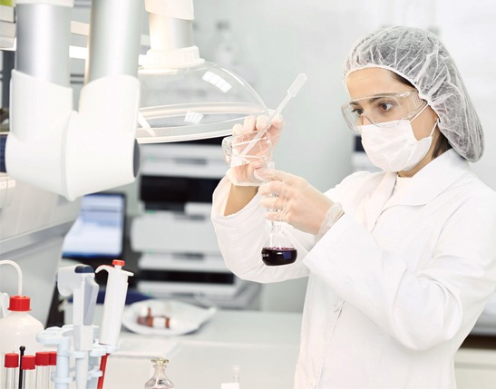
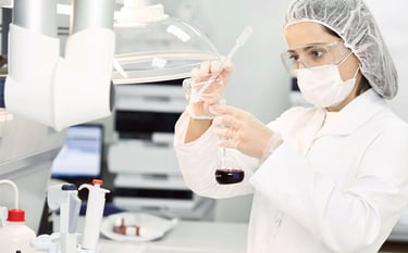
Somos creadores del Medio de Transporte Viral Virsenn, producto con innovación y desarrollo 100% mexicano
Laboratorios CIDSA
Calidad de alta competencia
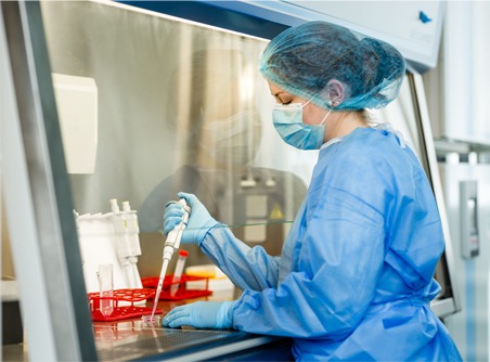
Laboratorio 100% Mexicano que ha hecho de la calidad un compromiso. No solo es por lo que trabajamos - es nuestro principio y responsabilidad.
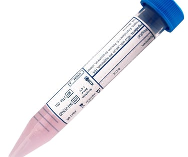
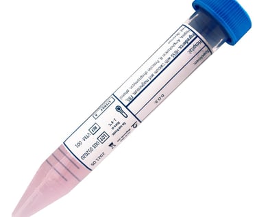
Desarrollamos el Medio de Transporte Viral (MTV) de acuerdo a sus especificaciones
Nuestro laboratorio además de ofrecer el Medio deTransporte Viral estandar, también nos adecuamos a sus necesidades. Usted puede solicitar el MTV de acuerdo a la formula que usted requiera.

Experiencia
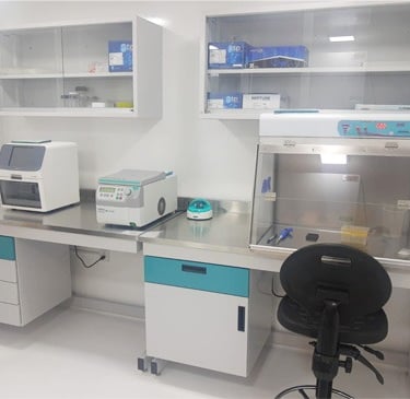
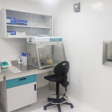
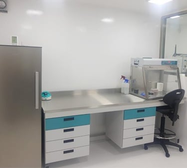
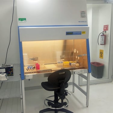
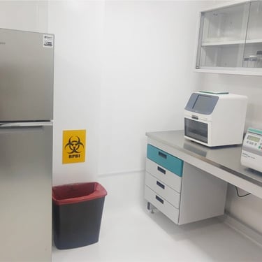
Somos un laboratorio privado de recién creación y con una experiencia en Ciencias de la Vida de más de 40 años. Nuestras instalaciones cuentan con certificaciones para realizar pruebas moleculares y diagnóstico bajo altos estándares de calidad y reproducibilidad, con personal entrenado y capacitado en Biología Molecular y en prácticas BSL2. Seguimos protocolos y kits validados por el InDRE..
Contacto
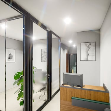
Laboratorios y Oficinas
• Matriz y Laboratorios en Queretaro Libramiento Norponiente
• Laboratorios en Zacatecas - Col. Bonito Pueblo, Guadalupe Zac. - Col. Spauaz, Guadalupe Zac.
• Oficinas y Almacen en CDMX
Información
Tel. 55-8961-0759 CDMX www.cidsamexico.com ventas@cidsamexico.com
Tel. Zacatecas 492-921-9474 WhatsApp 55-3621-4100