
iFluor® 350 SE es razonablemente estable y muestra buena reactividad y selectividad con los grupos amino de proteínas.
Descripción
Los tintes iFluor® de AAT Bioquest están optimizados para marcar proteínas, en particular, anticuerpos. Estos tintes son brillantes, fotoestables y tienen un enfriamiento mínimo de las proteínas. Pueden excitarse bien con las principales líneas láser de los instrumentos de fluorescencia (ejem. 350, 405, 488, 555 y 633 nm).
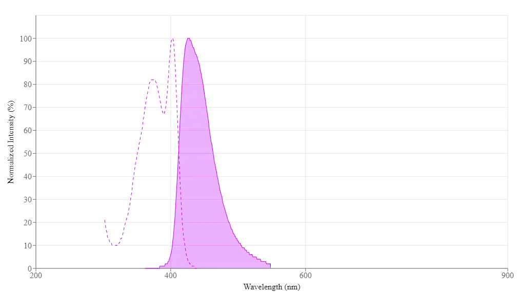
Los tintes iFluor® 405 tienen un máximo de excitación y emisión de fluorescencia de ~406 nm y ~427 nm respectivamente. Estas características espectrales los convierten en una excelente alternativa al colorante de etiquetado AMCA y Alexa Fluor® 405 (Alexa Fluor® es la marca registrada de Invitrogen).
iFluor® 405 SE es razonablemente estable y muestra buena reactividad y selectividad con los grupos amino de proteínas.
| Catalogo | Producto | Presentación |
|---|---|---|
| AAT-1021 | iFluor® 405 succinimidyl ester | 1mg |
| AAT-71021 | iFluor® 405 succinimidyl ester | 100 ug |
| AAT-71501 | iFluor® 405 succinimidyl ester | 5mg |
| AAT-71551 | iFluor® 405 succinimidyl ester | 10mg |
Importante: Solo para uso en investigación (RUO). Almacenamiento: Congelación (< -15 °C). Minimizar la exposición a la luz.
Propiedades fisicas
| Peso Molecular | 755.58 |
| Disolvente | DMSO |
Espectro
Abrir en Advanced Spectrum Viewer
Propiedades espectrales
| Factor de corrección (260 nm) | 0.48 |
| Factor de corrección (280 nm) | 0.77 |
| Coeficiente de extinción (cm -1 M -1) | 370001 |
| Excitación (nm) | 403 |
| Emisión (nm) | 427 |
| Rendimiento cuántico | 0.911 |
Calculadora
Preparación de la solución de stock común
Volumen de DMSO necesario para reconstituir la masa específica de succinimidil éster iFluor® 405 a la concentración dada. Tenga en cuenta que el volumen es solo para preparar la solución madre. Consulte el protocolo experimental de la muestra para conocer los buffers experimentales/fisiológicos apropiados.
| 0.1 mg | 0.5 mg | 1 mg | 5 mg | 10 mg | |
| 1 mM | 132.349 µL | 661.743 µL | 1.323 mL | 6.617 mL | 13.235 mL |
| 5 mM | 26.47 µL | 132.349 µL | 264.697 µL | 1.323 mL | 2.647 mL |
| 10 mM | 13.235 µL | 66.174 µL | 132.349 µL | 661.743 µL | 1.323 mL |
Imagenes

Figura 1. Las células HL-60 se incubaron con (rojo, +) o sin (verde, -) anti-HLA-ABC humano (W6/32 mAb), seguido de iFluor® 405 cabra anti-ratón IgG (H&L). La señal de fluorescencia se controló utilizando un citómetro de flujo ACEA NovoCyte en el canal Pacific Blue (Ex/Em=405/445 nm).
Productos Similares
| Name | Excitation (nm) | Emission (nm) | Extinction coefficient (cm -1 M -1) | Quantum yield | Correction Factor (260 nm) | Correction Factor (280 nm) |
| iFluor® 350 succinimidyl ester | 345 | 450 | 200001 | 0.951 | 0.83 | 0.23 |
| iFluor® 488 succinimidyl ester | 491 | 516 | 750001 | 0.91 | 0.21 | 0.11 |
| iFluor® 514 succinimidyl ester | 511 | 527 | 750001 | 0.831 | 0.265 | 0.116 |
| iFluor® 532 succinimidyl ester | 537 | 560 | 900001 | 0.681 | 0.26 | 0.16 |
| iFluor® 555 succinimidyl ester | 557 | 570 | 1000001 | 0.641 | 0.23 | 0.14 |
| iFluor® 594 succinimidyl ester | 588 | 604 | 1800001 | 0.531 | 0.05 | 0.04 |
| iFluor® 633 succinimidyl ester | 640 | 654 | 2500001 | 0.291 | 0.062 | 0.044 |
| iFluor® 647 succinimidyl ester | 656 | 670 | 2500001 | 0.251 | 0.03 | 0.03 |
| iFluor® 660 succinimidyl ester | 663 | 678 | 2500001 | 0.261 | 0.07 | 0.08 |
Bibliografía
Arabidopsis cryptochrome 2 forms photobodies with TCP22 under blue light and regulates the circadian clock
Authors: Mo, Weiliang and Zhang, Junchuan and Zhang, Li and Yang, Zhenming and Yang, Liang and Yao, Nan and Xiao, Yong and Li, Tianhong and Li, Yaxing and Zhang, Guangmei and others,
Journal: Nature communications (2022): 1–15
Arc weakens synapses by dispersing AMPA receptors from postsynaptic density via modulating PSD phase separation
Authors: Chen, Xudong and Jia, Bowen and Araki, Yoichi and Liu, Bian and Ye, Fei and Huganir, Richard and Zhang, Mingjie
Journal: Cell Research (2022): 914–930
Kindlin2-mediated phase separation underlies integrin adhesion formation
Authors: Li, Yujie and Zhang, Ting and Li, Huadong and Yang, Haibin and Lin, Ruihong and Sun, Kang and Wang, Lei and Zhang, Jing and Wei, Zhiyi and Yu, Cong
Journal: BioRxiv (2020)
CaMKII activation triggers persistent formation and segregation of postsynaptic liquid phase
Authors: Hosokawa, Tomohisa and Liu, Pin-Wu and Cai, Qixu and Ferreira, Joana S and Levet, Florian and Butler, Corey and Sibarita, Jean-Baptiste and Choquet, Daniel and Groc, Laurent and Hosy, Eric and others,
Journal: bioRxiv (2020)
Par complex cluster formation mediated by phase separation
Authors: Liu, Ziheng and Yang, Ying and Gu, Aihong and Xu, Jiawen and Mao, Ying and Lu, Haojie and Hu, Weiguo and Lei, Qun-Ying and Li, Zhouhua and Zhang, Mingjie and others,
Journal: Nature communications (2020): 1–18
Phase separation-mediated TARP/MAGUK complex condensation and AMPA receptor synaptic transmission
Authors: Zeng, Menglong and D{\’\i}az-Alonso, Javier and Ye, Fei and Chen, Xudong and Xu, Jia and Ji, Zeyang and Nicoll, Roger A and Zhang, Mingjie
Journal: Neuron (2019): 529–543
Reconstituted postsynaptic density as a molecular platform for understanding synapse formation and plasticity
Authors: Zeng, Menglong and Chen, Xudong and Guan, Dongshi and Xu, Jia and Wu, Haowei and Tong, Penger and Zhang, Mingjie
Journal: Cell (2018): 1172–1187
Deep Sequencing Analysis of the Eha-Regulated Transcriptome of Edwardsiella tarda Following Acidification
Authors: Gao, D and Liu, N and Li, Y and Zhang, Y and Liu, G and others, undefined
Journal: Metabolomics (Los Angel) (2017): 2153–0769
Suramin inhibits cullin-RING E3 ubiquitin ligases
Authors: Wu, Kenneth and Chong, Robert A and Yu, Qing and Bai, Jin and Spratt, Donald E and Ching, Kevin and Lee, Chan and Miao, Haibin and Tappin, Inger and Hurwitz, Jerard and others, undefined
Journal: Proceedings of the National Academy of Sciences (2016): E2011–E2018
Glycosaminoglycan mimicry by COAM reduces melanoma growth through chemokine induction and function
Authors: Piccard, Helene and Berghmans, Nele and Korpos, Eva and Dillen, Chris and Aelst, Ilse Van and Li, S and ra , undefined and Martens, Erik and Liekens, S and ra , undefined and Noppen, Sam and Damme, Jo Van and others, undefined
Journal: International Journal of Cancer (2012): E425–E436
Referencias
Ver todas las 49 referencias: Citation Explorer
Sequential ordering among multicolor fluorophores for protein labeling facility via aggregation-elimination based beta-lactam probes
Authors: Sadhu KK, Mizukami S, Watanabe S, Kikuchi K.
Journal: Mol Biosyst (2011): 1766
Visualizing dengue virus through Alexa Fluor labeling
Authors: Zhang S, Tan HC, Ooi EE.
Journal: J Vis Exp. (2011)
Fluorescent “Turn-on” system utilizing a quencher-conjugated peptide for specific protein labeling of living cells
Authors: Arai S, Yoon SI, Murata A, Takabayashi M, Wu X, Lu Y, Takeoka S, Ozaki M.
Journal: Biochem Biophys Res Commun (2011): 211
Neuroanatomical basis of clinical joint application of “Jinggu” (BL 64, a source-acupoint) and “Dazhong” (KI 4, a Luo-acupoint) in the rat: a double-labeling study of cholera toxin subunit B conjugated with Alexa Fluor 488 and 594
Authors: Cui JJ, Zhu XL, Ji CF, Jing XH, Bai WZ.
Journal: Zhen Ci Yan Jiu (2011): 262
Simultaneous detection of virulence factors from a colony in diarrheagenic Escherichia coli by a multiplex PCR assay with Alexa Fluor-labeled primers
Authors: Kuwayama M, Shigemoto N, Oohara S, Tanizawa Y, Yamada H, Takeda Y, Matsuo T, Fukuda S.
Journal: J Microbiol Methods (2011): 119
Alexa Fluor 546-ArIB[V11L;V16A] is a potent ligand for selectively labeling alpha 7 nicotinic acetylcholine receptors
Authors: Hone AJ, Whiteaker P, Mohn JL, Jacob MH, McIntosh JM.
Journal: J Neurochem (2010): 994
Asymmetric trimethine 3H-indocyanine dyes: efficient synthesis and protein labeling
Authors: Song F, Wang L, Qiao X, Wang B, Sun S, Fan J, Zhang L, Peng X.
Journal: Org Biomol Chem (2010): 4249
Neuroanatomical characteristics of acupoint “Chengshan” (BL 57) in the rat: a cholera toxin subunit B conjugated with Alexa Fluor 488 method study
Authors: Zhu XL, Bai WZ, Wu FD, Jiang J, Jing XH.
Journal: Zhen Ci Yan Jiu (2010): 433
Photoactivatable and photoconvertible fluorescent probes for protein labeling
Authors: Maurel D, Banala S, Laroche T, Johnsson K.
Journal: ACS Chem Biol (2010): 507
Novel Alexa Fluor-488 labeled antagonist of the A(2A) adenosine receptor: Application to a fluorescence polarization-based receptor binding assay
Authors: Kecskes M, Kumar TS, Yoo L, Gao ZG, Jacobson KA.
Journal: Biochem Pharmacol (2010): 506
Application Notes
iFluor® Dye Selection Guide
A New Protein Crosslinking Method for Labeling and Modifying Antibodies
Abbreviation of Common Chemical Compounds Related to Peptides
Bright Tide Fluor™-Based Fluorescent Peptides and Their Applications In Drug Discovery and Disease Diagnosis
FITC (Fluorescein isothiocyanate)
FAQ
What are common laser lines used in flow cytometry?
What are the spectral properties of iFluor dyes?
Are any of the cyanine dyes infrared?
Are coumarin dyes pH sensitive?
Are there any alternatives to BrdU (Bromodeoxyuridine)?
AssayWise
A practical guide for use of PE and APC in flow cytometry
Calbryte™ series now available
Buccutite™ Fluorescent Protein and Tandem Dye Antibody Labeling Kits
Fundamentals of Flow Cytometry
ReadiUse™ Lyophilized Phycobiliproteins