La maleimida iFluor® 860 es reactiva con tiol y se puede usar fácilmente para conjugar biomoléculas que contienen tiol.
Descripción
Las imágenes de fluorescencia in vivo utilizan una cámara sensible para detectar la emisión de fluorescencia de los fluoróforos en pequeños animales vivos de cuerpo entero. Para superar la atenuación de fotones en el tejido vivo, generalmente se prefieren los fluoróforos con emisión prolongada en la región infrarroja (IR). Los avances recientes en las estrategias de obtención de imágenes y las técnicas de reportero para la obtención de imágenes por fluorescencia in vivo incluyen enfoques novedosos para mejorar la especificidad y la afinidad de las sondas y para modular y amplificar la señal en los sitios objetivo para aumentar la sensibilidad. Otros desarrollos emergentes tienen como objetivo lograr imágenes de fluorescencia in vivo de alta resolución, multimodalidad y basadas en la vida útil.
Nuestro iFluor® 860 está diseñado para marcar proteínas y otras biomoléculas con fluorescencia infrarroja. Los conjugados preparados con iFluor® 860 tienen excitación y emisión en el rango IR. La emisión del colorante iFluor® 860 está bien separada de los fluoróforos de rojo lejano comúnmente utilizados, como Cy5, Cy7 o la aloficocianina (APC), lo que facilita el análisis multicolor. Este fluoróforo también es útil para aplicaciones de imágenes in vivo de animales pequeños u otras aplicaciones de imágenes que requieren detección IR. La maleimida iFluor® 860 es reactiva con tiol y se puede usar fácilmente para conjugar biomoléculas que contienen tiol.
| Catalogo | Producto | Presentación |
|---|---|---|
| AAT-1408 | iFluor® 860 maleimide | 1 mg |
Importante: Solo para uso en investigación (RUO). Almacenamiento: Congelación (< -15 °C). Minimizar la exposición a la luz.
Propiedades fisicas
| Peso Molecular | 1647.66 |
| Disolvente | DMSO |
Espectro
Abrir en Advanced Spectrum Viewer

Propiedades espectrales
| Factor de corrección (260 nm) | 0.1 |
| Factor de corrección (280 nm) | 0.14 |
| Coeficiente de extinción (cm -1 M -1) | 2500001 |
| Excitación (nm) | 853 |
| Emisión (nm) | 878 |
Calculadora
Preparación de la solución de stock común
Volumen de DMSO necesario para reconstituir la masa específica de iFluor® 860 maleimida a la concentración dada. Tenga en cuenta que el volumen es solo para preparar la solución madre. Consulte el protocolo experimental de muestra para conocer los buffers experimentales/fisiológicos apropiados.
| 0.1mg | 0.5mg | 1mg | 5mg | 10mg | |
| 1 mM | 60.692 µL | 303.461 µL | 606.921 µL | 3.035 mL | 6.069 mL |
| 5 mM | 12.138 µL | 60.692 µL | 121.384 µL | 606.921 µL | 1.214 mL |
| 10 mM | 6.069 µL | 30.346 µL | 60.692 µL | 303.461 µL | 606.921 µL |
Imagenes

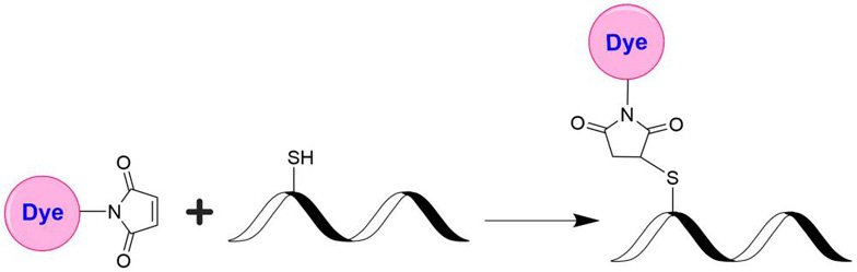
Figura 1. Las maleimidas de colorantes fluorescentes (p. ej., iFluor 860 maleimida) son la herramienta más popular para conjugar colorantes con un péptido, proteína, anticuerpo, oligonucleótido modificado con tiol o ácido nucleico a través de su grupo SH. Las maleimidas reaccionan rápidamente con el grupo tiol de las proteínas, los oligonucleótidos modificados con tiol y otras moléculas que contienen tiol en condiciones neutras. Los conjugados de tinte resultantes son bastante estables.
Productos Similares
| Name | Excitation (nm) | Emission (nm) | Extinction coefficient (cm -1 M -1) | Quantum yield | Correction Factor (260 nm) | Correction Factor (280 nm) |
| iFluor® 350 maleimide | 345 | 450 | 200001 | 0.951 | 0.83 | 0.23 |
| iFluor® 488 maleimide | 491 | 516 | 750001 | 0.91 | 0.21 | 0.11 |
| iFluor® 555 maleimide | 557 | 570 | 1000001 | 0.641 | 0.23 | 0.14 |
| iFluor® 647 maleimide | 656 | 670 | 2500001 | 0.251 | 0.03 | 0.03 |
| iFluor® 680 maleimide | 684 | 701 | 2200001 | 0.231 | 0.097 | 0.094 |
| iFluor® 700 maleimide | 690 | 713 | 2200001 | 0.231 | 0.09 | 0.04 |
| iFluor® 750 maleimide | 757 | 779 | 2750001 | 0.121 | 0.044 | 0.039 |
| iFluor® 790 maleimide | 787 | 812 | 2500001 | 0.131 | 0.1 | 0.09 |
| iFluor® 800 maleimide | 801 | 820 | 2500001 | 0.111 | 0.03 | 0.08 |
Bibliografía
Pancreatic Cancer Cells Undergo Immunogenic Cell Death upon Exposure to Gas Plasma-Oxidized Ringer’s Lactate
Authors: Miebach, Lea and Mohamed, Hager and Wende, Kristian and Miller, Vandana and Bekeschus, Sander
Journal: Cancers (2023): 319
Identification of distinct functional thymic programming of fetal and pediatric human $\gamma$$\delta$ thymocytes via single-cell analysis
Authors: Sanchez Sanchez, Guillem and Papadopoulou, Maria and Azouz, Abdulkader and Tafesse, Yohannes and Mishra, Archita and Chan, Jerry KY and Fan, Yiping and Verdebout, Isoline and Porco, Silvana and Libert, Fr{\’e}d{\’e}rick and others,
Journal: Nature communications (2022): 1–19
Conductive Gas Plasma Treatment Augments Tumor Toxicity of Ringer’s Lactate Solutions in a Model of Peritoneal Carcinomatosis
Authors: Miebach, Lea and Freund, Eric and Cecchini, Alessandra Louren{\c{c}}o and Bekeschus, Sander
Journal: Antioxidants (2022): 1439
A versatile platform for generating engineered extracellular vesicles with defined therapeutic properties
Authors: Dooley, Kevin and McConnell, Russell E and Xu, Ke and Lewis, Nuruddeen D and Haupt, Sonya and Youniss, Madeleine R and Martin, Shelly and Sia, Chang Ling and McCoy, Christine and Moniz, Raymond J and others,
Journal: Molecular Therapy (2021): 1729–1743
Soluble ST2 links inflammation to outcome after subarachnoid hemorrhage
Authors: Bevers, Matthew B and Wolcott, Zoe and Bache, Søren and Hansen, Christina and Sastre, Cristina and Mylvaganam, Ravi and Koch, Matthew J and Patel, Aman B and Møller, Kirsten and Kimberly, W Taylor
Journal: Annals of neurology (2019)
Nanovesicle delivery to the liver via retinol binding protein and platelet-derived growth factor receptors: how targeting ligands affect biodistribution
Authors: Hsu, Ching-Yun and Chen, Chun-Han and Aljuffali, Ibrahim A and Dai, You-Shan and Fang, Jia-You
Journal: Nanomedicine (2017)
18–Color Human Blood Phenotyping Made Easy with Flow Cytometry
Authors: McCracken, James and Lawson, Jonel
Referencias
Ver todas las 18 referencias: Citation Explorer
A target cell-specific activatable fluorescence probe for in vivo molecular imaging of cancer based on a self-quenched avidin-rhodamine conjugate
Authors: Hama Y, Urano Y, Koyama Y, Kamiya M, Bernardo M, Paik RS, Shin IS, Paik CH, Choyke PL, Kobayashi H.
Journal: Cancer Res (2007): 2791
Fluorescence imaging in vivo: recent advances
Authors: Rao J, Dragulescu-Andrasi A, Yao H.
Journal: Curr Opin Biotechnol (2007): 17
Ex vivo fluorescence imaging of normal and malignant urothelial cells to enhance early diagnosis
Authors: Steenkeste K, Lecart S, Deniset A, Pernot P, Eschwege P, Ferlicot S, Leveque-Fort S, Bri and et R, Fontaine-Aupart MP.
Journal: Photochem Photobiol (2007): 1157
In vivo monitoring the fate of Cy5.5-Tat labeled T lymphocytes by quantitative near-infrared fluorescence imaging during acute brain inflammation in a rat model of experimental autoimmune encephalomyelitis
Authors: Berger C, Gremlich HU, Schmidt P, Cannet C, Kneuer R, Hiest and P, Rausch M, Rudin M.
Journal: J Immunol Methods (2007): 65
A protocol for imaging alternative splicing regulation in vivo using fluorescence reporters in transgenic mice
Authors: Bonano VI, Oltean S, Garcia-Blanco MA.
Journal: Nat Protoc (2007): 2166
In vivo imaging of the bronchial wall microstructure using fibered confocal fluorescence microscopy
Authors: Thiberville L, Moreno-Swirc S, Vercauteren T, Peltier E, Cave C, Bourg Heckly G.
Journal: Am J Respir Crit Care Med (2007): 22
In Vivo Fluorescence Microscopic Imaging for Dynamic Quantitative Assessment of Intestinal Mucosa Permeability in Mice
Authors: Szabo A, Vollmar B, Boros M, Menger MD.
Journal: J Surg Res. (2007)
In vivo spectral fluorescence imaging of submillimeter peritoneal cancer implants using a lectin-targeted optical agent
Authors: Hama Y, Urano Y, Koyama Y, Kamiya M, Bernardo M, Paik RS, Krishna MC, Choyke PL, Kobayashi H.
Journal: Neoplasia (2006): 607
In vivo imaging of green fluorescent protein-expressing cells in transgenic animals using fibred confocal fluorescence microscopy
Authors: Al-Gubory KH, Houdebine LM.
Journal: Eur J Cell Biol (2006): 837
In vivo near-infrared fluorescence imaging of integrin alphavbeta3 in an orthotopic glioblastoma model
Authors: Hsu AR, Hou LC, Veeravagu A, Greve JM, Vogel H, Tse V, Chen X.
Journal: Mol Imaging Biol (2006): 315
Guia de Selección tintes iFlour®