Kit de seguimiento de células vivas reparable Cell Explorer™, en particular está diseñado para etiquetar uniformemente células vivas en fluorescencia roja para los estudios que requieren que las moléculas de etiquetas fluorescentes se retengan dentro de las células durante un tiempo relativamente más largo.
Descripción
Kit de seguimiento de células vivas reparable Cell Explorer™ *fluorescencia roja*
Nuestros kits de imágenes de fluorescencia Cell Explorer™ son un conjunto de herramientas para etiquetar células para investigaciones microscópicas de fluorescencia de funciones celulares. El etiquetado eficaz de las células proporciona un método poderoso para estudiar eventos celulares en un contexto espacial y temporal.
Este kit en particular está diseñado para etiquetar uniformemente células vivas en fluorescencia roja para los estudios que requieren que las moléculas de etiquetas fluorescentes se retengan dentro de las células durante un tiempo relativamente más largo. Las células se pueden fijar para conservar el patrón de imagen.
El kit utiliza un tinte no fluorescente que lleva una fracción que retiene las células. Se vuelve fuertemente fluorescente al entrar en las células vivas y queda atrapado dentro de las células vivas para dar una señal de fluorescencia estable durante un tiempo relativamente largo. Este producto es un compuesto hidrofóbico que penetra fácilmente en las células vivas intactas.
El proceso de etiquetado es sólido y requiere un tiempo mínimo de intervención. Puede adaptarse fácilmente a una amplia variedad de plataformas de fluorescencia, como ensayos de microplacas, inmunocitoquímica y citometría de flujo.
Es útil para una variedad de estudios, incluida la adhesión celular, la quimiotaxis, la resistencia a múltiples fármacos, la viabilidad celular, la apoptosis y la citotoxicidad. El kit proporciona todos los componentes esenciales con un protocolo de etiquetado celular optimizado.
Nombre en Ingles: Cell Explorer™ Fixable Live Cell Tracking Kit *Red Fluorescence*
| Catalogo | Producto | Presentación |
|---|---|---|
| AAT-22625 | Kit de seguimiento de células vivas reparable Cell Explorer™ | 200 pruebas |
Importante: Solo para uso en investigación (RUO). Almacenamiento a <-15 °C. Minimizar exposición a la luz.
Componentes
| Component A: Track It™ Red | 1 vial |
| Component B: Assay Buffer | 1 frasco (20 mL) |
| Component C: DMSO | 1 vial (100 µL) |
Plataforma
Citómetro de Flujo
| Excitación | 488 nm laser |
| Emisión | 610/20 nm filtro |
| Epecificaciones Instrumento | Canal PE-Texas Red |
Microscopio de Fluorescencia
| Excitación | 570 nm |
| Emisión | 600 nm |
| Placa Recomendada | Pared negra/fondo claro |
| Especificaciones Instrumento | Filtro Texas Red |
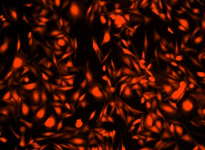
Imagen
Figura 1.
Imagen de células HeLa teñidas con el kit de seguimiento de células vivas Cell Explorer™ en una placa de 96 pocillos de pared negra/fondo transparente Costar. Las células se tiñeron con Track It™ Red (Cat. 22625) y las imágenes se adquirieron utilizando un microscopio de fluorescencia con un juego de filtros Texas Red.
Formato Alternativo
Productos Relacionados
Bibliografia
The Biological Effects of Interleukin-17A on Adhesion Molecules Expression and Foam Cell Formation in Atherosclerotic Lesions
Authors: Shiotsugu, Shohei and Okinaga, Toshinori and Habu, Manabu and Yoshiga, Daigo and Yoshioka, Izumi and Nishihara, Tatsuji and Ariyoshi, Wataru
Journal: Journal of Interferon & Cytokine Research (2019)
Autophagy proteins are not universally required for phagosome maturation
Authors: Cemma, Marija and Grinstein, Sergio and Brumell, John H
Journal: Autophagy (2016): 1440–1446
Differential detection of tumor cells using a combination of cell rolling, multivalent binding, and multiple antibodies
Authors: Myung, Ja Hye and Gajjar, Khyati A and Chen, Jihua and Molokie, Robert E and Hong, Seungpyo
Journal: Analytical chemistry (2014): 6088–6094
Versatile fabrication of nanoscale sol–gel bioactive glass particles for efficient bone tissue regeneration
Authors: Lei, Bo and Chen, Xiaofeng and Han, Xue and Zhou, Jiaan
Journal: Journal of Materials Chemistry (2012): 16906–16913
Referencias
Ver todas las 26 referencias: Citation Explorer
Requirements, features, and performance of high content screening platforms
Authors: Gough AH, Johnston PA.
Journal: Methods Mol Biol (2007): 41
A pharmaceutical company user’s perspective on the potential of high content screening in drug discovery
Authors: Hoffman AF, Garippa RJ.
Journal: Methods Mol Biol (2007): 19
Optimizing the integration of immunoreagents and fluorescent probes for multiplexed high content screening assays
Authors: Giuliano KA., undefined
Journal: Methods Mol Biol (2007): 189
Past, present, and future of high content screening and the field of cellomics
Authors: Taylor DL., undefined
Journal: Methods Mol Biol (2007): 3
High-content fluorescence-based screening for epigenetic modulators
Authors: Martinez ED, Dull AB, Beutler JA, Hager GL.
Journal: Methods Enzymol (2006): 21
Application of laser-scanning fluorescence microplate cytometry in high content screening
Authors: Bowen WP, Wylie PG.
Journal: Assay Drug Dev Technol (2006): 209
High-content screening of known G protein-coupled receptors by arrestin translocation
Authors: Hudson CC, Oakley RH, Sjaastad MD, Loomis CR.
Journal: Methods Enzymol (2006): 63
Evaluation of a high-content screening fluorescence-based assay analyzing the pharmacological modulation of lipid homeostasis in human macrophages
Authors: Werner T, Liebisch G, Gr and l M, Schmitz G.
Journal: Cytometry A (2006): 200
Automated high content screening for phosphoinositide 3 kinase inhibition using an AKT 1 redistribution assay
Authors: Wolff M, Haasen D, Merk S, Kroner M, Maier U, Bordel S, Wiedenmann J, Nienhaus GU, Valler M, Heilker R.
Journal: Comb Chem High Throughput Screen (2006): 339
High concordance of drug-induced human hepatotoxicity with in vitro cytotoxicity measured in a novel cell-based model using high content screening
Authors: O’Brien P J, Irwin W, Diaz D, Howard-Cofield E, Krejsa CM, Slaughter MR, Gao B, Kaludercic N, Angeline A, Bernardi P, Brain P, Hougham C.
Journal: Arch Toxicol (2006): 580
Application Notes
A New Protein Crosslinking Method for Labeling and Modifying Antibodies
A Novel Fluorescent Probe for Imaging and Detecting Hydroxyl Radical in Living Cells
Abbreviation of Common Chemical Compounds Related to Peptides
Annexin V
Buccutite™ Bioconjugation Technology
FAQ
Do you have fixable live cell staining assay kits?
What is the number of passages your Cell Explorer Live Cell Tracking assays can undergo?
Are there any alternatives to BrdU (Bromodeoxyuridine)?
Are there any alternatives to Cy5?
Can 7-AAD be fixed?
AssayWise
HRP Antibody Labeling Using Buccutite™ Crosslinking Technology
iFluor® 700 Dyes
Hydroxyl Radical Detection
Buccutite™ Conjugation Kits: Quick and Easy Antibody Labeling
Nucleic Acid Detection, Quantification and Imaging