La R-Ficoeritrina (PE) se aísla de algas rojas. Su pico de absorción principal está a 565 nm con picos secundarios a 496 y 545 nm.
Descripción
La R-Ficoeritrina (PE) se aísla de algas rojas. Su pico de absorción principal está a 565 nm con picos secundarios a 496 y 545 nm. La prominencia relativa de los picos secundarios varía significativamente entre los R-PE de diferentes especies. PE tiene tres tipos de subunidades: alfa (20.000 daltons), beta (20.000 daltons) y gamma (30.000 daltons). Se ha encontrado que el peso molecular del PE intacto es de aproximadamente 240.000 daltons. La subunidad alfa de PE contiene solo el cromóforo ficoeritrobilina (PEB), mientras que las subunidades beta y gamma contienen tanto PEB como ficourobilina (PUB). La variabilidad en los espectros de absorción de PE de varias especies refleja diferencias en la relación PEB/PUB de las subunidades. La PE y la B-PE estrechamente relacionada son las ficobiliproteínas más intensamente fluorescentes, con eficiencias cuánticas probablemente superiores al 90 %, y su fluorescencia naranja es fácilmente visible a simple vista en cualquier solución moderadamente concentrada.
| Catalogo | Producto | Presentación |
|---|---|---|
| AAT-2558 | PE [R-Phycoerythrin] *CAS 11016-17-4* | 1 mg |
| AAT-2556 | PE [R-Phycoerythrin] *CAS 11016-17-4* | 10 mg |
| AAT-2557 | PE [R-Phycoerythrin] *CAS 11016-17-4* | 100 mg |
Importante: Solo para uso en investigación (RUO). Almacenamiento: Refrigeración (2-8 °C). Minimizar la exposición a la luz.
Propiedades fisicas
| Peso Molecular | -240000 |
| Disolvente | AGUA |
Espectro
Abrir en Advanced Spectrum Viewer
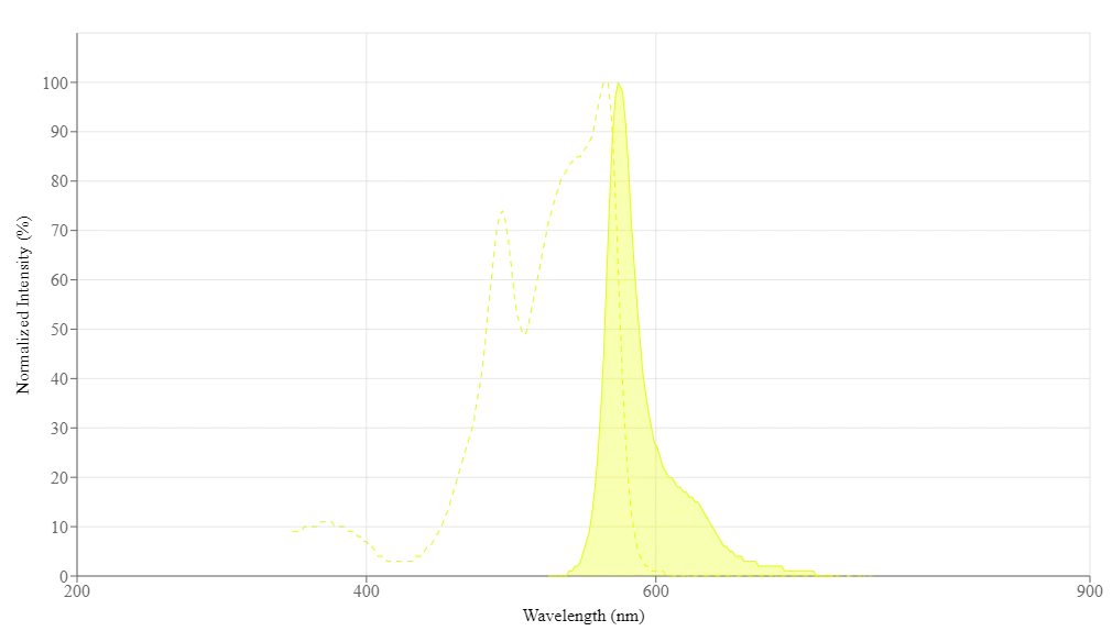
Propiedades espectrales
| Coeficiente de extinción (cm -1 M -1) | 1960000 |
| Excitación (nm) | 566 |
| Emisión (nm) | 574 |
| Rendimiento cuántico | 0.82 |
Imagenes
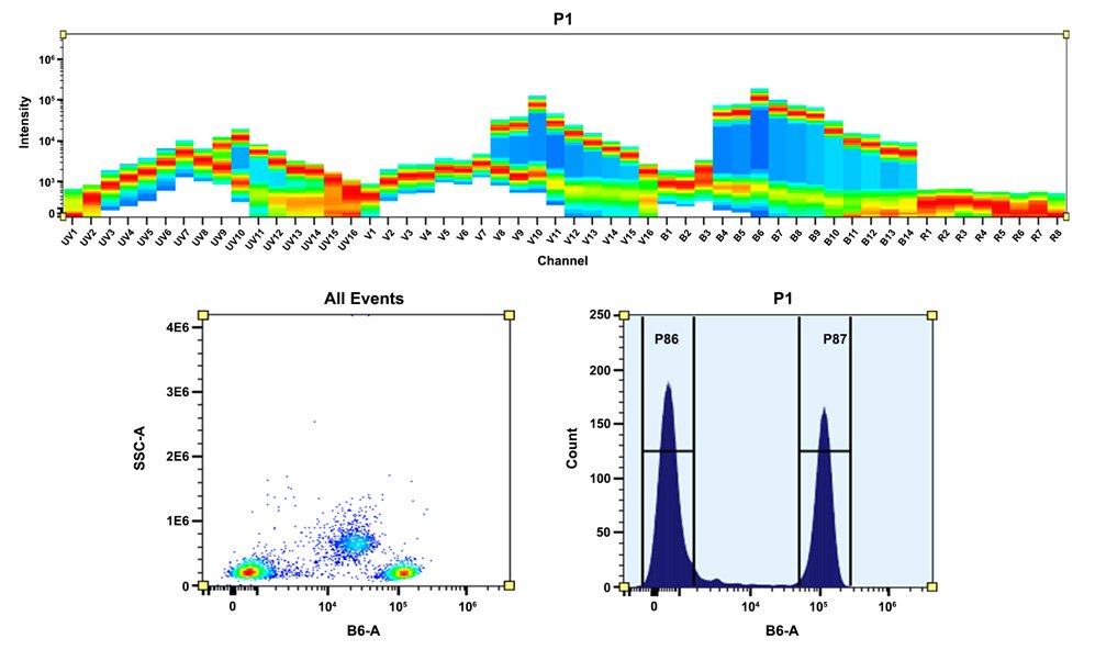
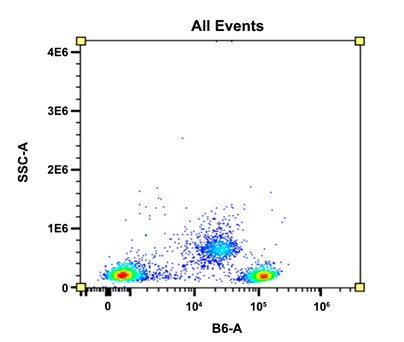
Figura 1. Arriba) El patrón espectral se generó utilizando un citómetro espectral de 4 láseres. Se utilizaron láseres desplazados espacialmente (355 nm, 405 nm, 488 nm y 640 nm) para crear cuatro perfiles de emisión distintos y luego, cuando se combinaron, produjeron la firma espectral general. Abajo) Análisis de citometría de flujo de PBMC teñidas con conjugado PE anti-CD4 humano *SK3*. La señal de fluorescencia se controló usando un citómetro de flujo Aurora en el canal B6-A específico de PE.
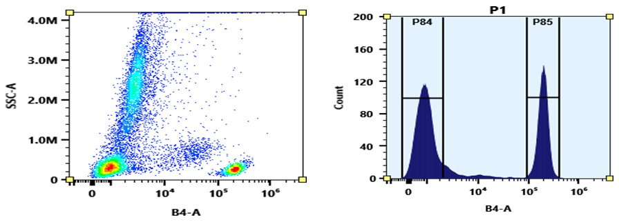
Figura 2. Análisis de citometría de flujo de sangre completa teñida con conjugado de PE anti-CD4 humano *SK3*. La señal de fluorescencia se controló utilizando un citómetro de flujo Aurora en el canal B4-A específico de PE.
Bibliografía
CD169+ subcapsular sinus macrophage-derived microvesicles are associated with light zone follicular dendritic cells
Authors: Chen, Xin and Zheng, Yuhan and Liu, Siming and Yu, Wenjing and Liu, Zhiduo
Journal: European Journal of Immunology (2022): 1581–1594
Nanobubbles Containing sPD-1 and Ce6 Mediate Combination Immunotherapy and Suppress Hepatocellular Carcinoma in Mice
Authors: Tan, Yandi and Yang, Shiqi and Ma, Yao and Li, Jinlin and Xie, Qian and Liu, Chaoqi and Zhao, Yun
Journal: International Journal of Nanomedicine (2021): 3241
Morphological Changes Induced By Extremely Low-Frequency Electric Fields
Authors: Imani, Mahdi and Kazemi, Sepide and Saviz, Mehrdad and Farahm, undefined and , Leila and Sadeghi, Behnam and Faraji-dana, Reza
Journal: Bioelectromagnetics (2019)
Referencias
Chromophore attachment to phycobiliprotein beta-subunits: phycocyanobilin:cysteine-beta84 phycobiliprotein lyase activity of CpeS-like protein from Anabaena Sp. PCC7120
Authors: Zhao KH, Su P, Li J, Tu JM, Zhou M, Bubenzer C, Scheer H.
Journal: J Biol Chem (2006): 8573
Excitation energy transfer from phycobiliprotein to chlorophyll d in intact cells of Acaryochloris marina studied by time- and wavelength-resolved fluorescence spectroscopy
Authors: Petrasek Z, Schmitt FJ, Theiss C, Huyer J, Chen M, Larkum A, Eichler HJ, Kemnitz K, Eckert HJ.
Journal: Photochem Photobiol Sci (2005): 1016
Single-molecule spectroscopy selectively probes donor and acceptor chromophores in the phycobiliprotein allophycocyanin
Authors: Loos D, Cotlet M, De Schryver F, Habuchi S, Hofkens J.
Journal: Biophys J (2004): 2598
Isolation and characterisation of phycobiliprotein rich mutant of cyanobacterium Synechocystis sp
Authors: Prasanna R, Dhar DW, Dominic TK, Tiwari ON, Singh PK.
Journal: Acta Biol Hung (2003): 113
Evaluation of Tolypothrix germplasm for phycobiliprotein content
Authors: Prasanna R, Prasanna BM, Mohammadi SA, Singh PK.
Journal: Folia Microbiol (Praha) (2003): 59
Co-ordinated expression of phycobiliprotein operons in the chromatically adapting cyanobacterium Calothrix PCC 7601: a role for RcaD and RcaG
Authors: Noubir S, Luque I, Ochoa de Alda JA, Perewoska I, T and eau de Marsac N, Cobley JG, Houmard J.
Journal: Mol Microbiol (2002): 749
Phycobiliprotein genes of the marine photosynthetic prokaryote Prochlorococcus: evidence for rapid evolution of genetic heterogeneity
Authors: Ting CS, Rocap G, King J, Chisholm SW.
Journal: Microbiology (2001): 3171
Phycobiliprotein-Fab conjugates as probes for single particle fluorescence imaging
Authors: Triantafilou K, Triantafilou M, Wilson KM.
Journal: Cytometry (2000): 226
Novel activity of a phycobiliprotein lyase: both the attachment of phycocyanobilin and the isomerization to phycoviolobilin are catalyzed by the proteins PecE and PecF encoded by the phycoerythrocyanin operon
Authors: Zhao KH, Deng MG, Zheng M, Zhou M, Parbel A, Storf M, Meyer M, Strohmann B, Scheer H.
Journal: FEBS Lett (2000): 9
Phycobiliprotein and fluorescence immunological assay
Authors: Wu P., undefined
Journal: Sheng Li Ke Xue Jin Zhan (2000): 82
Application Notes (en Ingles)
Phycobiliproteins and Their Fluorescent Labeling Applications
A New Protein Crosslinking Method for Labeling and Modifying Antibodies
Abbreviation of Common Chemical Compounds Related to Peptides